
Course Description
This course teaches the effects of mechanical forces (strains) on cells and how cells generate mechanical forces by their contraction. Critical to both topics is the mechanical behavior of the extracellular matrix (ECM), which the cells have synthesized and remodeled. The ECM affects cell behavior by mediating …
This course teaches the effects of mechanical forces (strains) on cells and how cells generate mechanical forces by their contraction. Critical to both topics is the mechanical behavior of the extracellular matrix (ECM), which the cells have synthesized and remodeled. The ECM affects cell behavior by mediating exogenous mechanical strains, which stimulate certain cellular processes.
Course Info
Instructors
Departments
Learning Resource Types
menu_book
Open Textbooks
notes
Lecture Notes
Readings
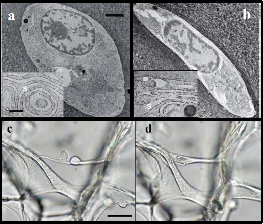
The figure shows 1) effects that mechanical forces can have on cells (a, b; chondrocytes), and 2) the generation of mechanical force by nonmuscle cells (c, d; chondrocytes).
- a. A chondrocyte in articular cartilage viewed by transmission electron microscopy, with its endoplasmic reticulum shown as s in the inset (scale bars, 3μm for a and b, and 400nm for the insets).
- b. A 50% compression of the cartilage sample results in a change in cell shape and compaction of the rough endoplasmic reticulum, responsible for the synthesis of extracellular matrix molecules. Alterations in the organelle can change the structure of the matrix molecules that the cell synthesizes. [Szafranski et al, Osteoarthr & Cart. 12:937 (2004)]
- c and d. Light microscopy images of chondrocytes, which other studies have shown express α-smooth muscle actin, seeded into a porous collagen-glycosaminoglycan scaffold, followed over a 5-hr period (scale bar, 50 μm for c and d). (c) For up to 40 min, the 2 chondrocytes on the horizontal strut have a rounded phenotype. (d) After 5 hrs, the cells spread on the strut and buckle it as a result of their contraction. The 40% shortening of the strut (from 144 μm in c to 89 μm in d) causes bending of the members to which it is attached, and contracture of the sponge-like scaffold. [Zaleskas et al, Biomat. 25:1299 (2004)]
(Figures a and b by Jon D. Szafranskiy, et al. and Figures c and d by Janice M. Zaleskas, et al. Copyright © Elsevier Ltd. Used with permission.)


